
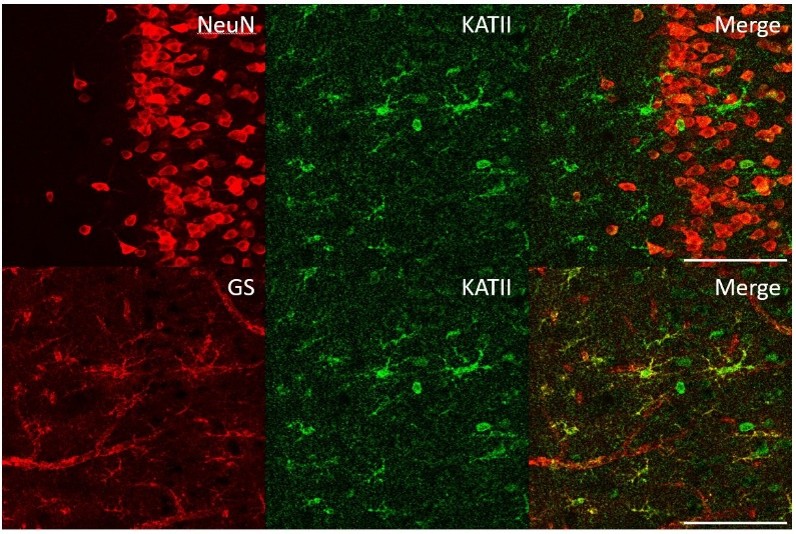
The brain is one of the most cell-type-diverse organs in the body. Identifying these cell types and mapping their spatial organization and interactions are essential to understanding their respective roles. This complexity poses a challenge for microscopy. A single brain section can contain neurons, astrocytes, microglia, oligodendrocytes, endothelial cells, and pericytes, each defined by its own combination of proteins and occupying a specific position relative to its neighbors.
For decades, immunohistochemistry and immunofluorescence have given neuroscientists a way to visualize individual proteins within this complex landscape. However, conventional single-marker or low-plex approaches fall short in the face of such cellular complexity. Understanding brain structure and disease increasingly requires researchers not only to identify individual proteins and cell types, but also to determine how they are spatially organized within the same tissue section. Multiplex imaging helps meet this need.
Multiplex imaging—the detection of multiple protein targets in the same sample—offers a way to build a more complete and spatially resolved view of the brain. Its value, however, depends on more than the number of markers included in a panel. Each target must also be detected with sufficient sensitivity and specificity to provide reliable biological information.
Applications in neuroscience
In basic neuroscience, common immunostaining targets include the neuronal marker NeuN, the astrocytic marker GFAP, the microglial marker Iba1, oligodendrocyte-lineage markers, and synaptic proteins such as synaptophysin, PSD-95, Bassoon, and Piccolo.
These markers allow researchers to investigate the cellular organization of brain regions such as the cortex, striatum, and corpus callosum. They can be used to examine neuronal density, glial distribution, synaptic architecture, and changes in protein expression across defined anatomical regions.
Neurodegenerative disease research has become one of the most active areas for multiplex imaging in neuroscience. In Alzheimer’s disease, for example, amyloid plaques form within a complex local environment containing neurons, astrocytes, microglia, and other cell types. Single-cell transcriptomic studies have shown that microglia surrounding plaques can adopt distinct disease-associated molecular states, but dissociated sequencing does not preserve where those cells were located in relation to plaques, neurons, or other tissue structures.
Spatial protein imaging can help translate these molecular signatures back into tissue context. Researchers can examine how microglial markers localize around individual amyloid plaques, how neuronal loss varies across the same region, and how astrocytic or immune responses relate to local pathology.
Related approaches are used to study tau-associated pathology in postmortem human tissue, changes in synaptic proteins, and glial and immune responses following localized brain injury. Multiplex immunofluorescence on formalin-fixed paraffin-embedded sections can also be used to examine how resident brain cells and infiltrating immune cells respond following therapeutic laser ablation in some cases of treatment-resistant epilepsy.
Why brain tissue is a distinct imaging challenge
Brain tissue presents demanding conditions for fluorescence imaging. Many biologically important targets—including low-abundance receptors, disease-associated proteins, and some synaptic markers—may sit close to the detection limit of standard indirect immunofluorescence.
At the same time, brain tissue can produce substantial fluorescence background. Aged and postmortem brain sections frequently contain lipofuscin, a pigment that accumulates in long-lived cells such as neurons and fluoresces broadly across the visible spectrum. Its emission can overlap with the fluorophores used to detect proteins of interest.
This background may be mistaken for genuine signal or may obscure weak staining. It therefore raises the effective detection threshold of the assay: abundant targets may remain readily visible, while lower-abundance markers become difficult to distinguish from autofluorescence.
Quenching steps, spectral unmixing, and computational background subtraction are consequently important considerations in neuroscience imaging workflows. These methods can reduce interference, but they do not eliminate the need to generate a sufficiently strong and specific signal at the target.
Sample preparation introduces additional challenges. Formalin-fixed paraffin-embedded brain tissue is widely used in archived human clinical and neuropathology collections, but fixation cross-links proteins, can mask epitopes, and may require aggressive antigen retrieval. Fresh-frozen sections may preserve some epitopes more effectively, but can be less resistant to repeated staining and elution cycles.
Whatever the sample type, the assay must preserve cortical layers, white and gray matter architecture, and fine neuronal and glial processes. Spatial context—not marker identity alone—is the information that researchers are ultimately trying to recover.
The sensitivity–multiplexing challenge
Sensitivity and multiplexing are often treated as separate assay characteristics, but in brain imaging they are tightly connected.
Signal-amplification methods can make low-abundance targets visible above tissue autofluorescence. However, strong amplification does not necessarily make it easy to detect many targets simultaneously. Some amplification chemistries are more naturally suited to single-marker or low-plex assays, while extending them to larger panels may require sequential processing.
Conversely, methods developed primarily to increase plex may introduce compromises in sensitivity. Cyclic multiplexing, for example, stains and images a small panel before removing or inactivating the detection reagents and repeating the process. This can enable dozens of markers to be accumulated across multiple rounds, but repeated staining, bleaching, stripping, or elution may progressively affect signal, epitopes, morphology, or tissue integrity.
Oligonucleotide-barcoded antibody systems use unique DNA sequences attached to antibodies so that successive cycles of fluorescent probe hybridization and removal can reveal small groups of markers. These methods reduce dependence on large numbers of spectrally distinct fluorophores, but they may still require multiple detection and imaging cycles.
The practical question is therefore not simply how many markers can be assigned to a panel. It is how many targets can be detected and quantified with sufficient sensitivity, specificity, and spatial fidelity on a single, often irreplaceable, tissue section.
A marker that cannot be distinguished reliably from background contributes little biological information, regardless of the nominal plex of the assay.
Combining signal amplification and multiplexing with MUSE®
One of the longest-established methods for increasing immunofluorescence sensitivity is tyramide signal amplification. It uses an enzyme-catalyzed reaction to deposit multiple reporter molecules around an antibody-bound target, increasing signal above tissue background. This can provide strong amplification and support sensitive low- to moderate-plex staining.
More recently, enzyme-free amplification methods based on programmable DNA nanotechnology have been developed. MUSE® belongs to this category and is designed to address sensitivity and multiplexing within the same assay architecture.
MUSE® uses programmable DNA assemblies to amplify and simultaneously detect multiple antibody-bound targets. The aim is not merely to increase signal or to increase plex independently, but to enable sensitive target detection (see Application Note) as multiple markers are combined in the same tissue section.
This combination is particularly relevant for brain imaging. A panel may need to include abundant cell-identity markers alongside weaker receptors, synaptic proteins, or disease-associated targets. Amplification helps bring these lower-abundance signals above the complex background of brain tissue, while multiplexing allows them to be interpreted in relation to surrounding cells, anatomical structures, and pathological features.
In this sense, amplification makes difficult targets visible, while multiplexing makes their spatial relationships interpretable.
How multiplex imaging expands neuroscience applications
Once markers can be detected with adequate sensitivity, multiplex imaging allows researchers to move beyond separate measurements of isolated targets.
A single panel can combine markers of neuronal, astrocytic, microglial, and oligodendrocyte identity with synaptic or disease-associated proteins. Rather than comparing adjacent sections stained independently, researchers can examine these features within the same tissue architecture.
This makes it possible to ask more relational biological questions:
- Which cell populations are located near a pathological structure?
- Which molecular states do those cells exhibit?
- How do synaptic features vary with anatomical location or local pathology?
- Which cellular responses occur together within the same microenvironment?
- How does protein co-expression differ across defined brain regions?
Such information cannot be obtained from single-marker staining alone. It is also complementary to dissociated single-cell sequencing, which can identify molecularly distinct cell populations but does not preserve their original positions or relationships within the tissue.
Multiplex imaging can therefore provide the spatial bridge between molecular cell states, anatomical organization, and disease pathology.
Looking ahead
Multiplex immunofluorescence imaging in neuroscience is increasingly converging with spatial transcriptomics, three-dimensional tissue clearing, and AI-based image segmentation. Together, these technologies are beginning to generate more comprehensive maps of brain cell types, connectivity, tissue architecture, and disease-associated changes.
The goal is not simply to detect the largest possible number of markers. It is to detect the largest possible number of biologically informative markers with sufficient sensitivity, specificity, and spatial fidelity.
As panels expand, the central challenge will be to preserve reliable detection of individual targets while connecting them to increasingly precise anatomical and functional context. Technologies that combine strong signal amplification with practical multiplexing will be important for achieving that balance.
Ultimately, it is the combination of sensitivity, breadth, and spatial resolution that will turn increasingly detailed images of the brain into a deeper mechanistic understanding of brain organization and neurological disease.